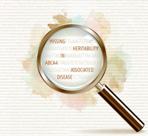
ABCA4-associated disease as a model for missing heritability in autosomal recessive disorders: novel noncoding splice, cis-regulatory, structural, and recurrent hypomorphic variants. Bauwens M, Garanto A, Sangermano R, Naessens S, Weisschuh N, De Zaeytijd J, Khan M2, Sadler F5, Balikova I, Van Cauwenbergh C1, Rosseel T, Bauwens J, De Leeneer K, De Jaegere S, Van Laethem T, De Vries M, Carss K, Arno G, Fakin A, Webster AR, de Ravel de l'Argentière TJL, Sznajer Y, Vuylsteke M, Kohl S, Wissinger B, Cherry T, Collin RWJ, Cremers FPM, Leroy BP, De Baere E. Genet Med. (2019). You can read the abstract below; for the full text click here.
Purpose: ABCA4-associated disease, a recessive retinal dystrophy, is hallmarked by a large proportion of patients with only one pathogenic ABCA4 variant, suggestive for missing heritability. Methods: By locus-specific analysis of ABCA4, combined with extensive functional studies, we aimed to unravel the missing alleles in a cohort of 67 patients (p), with one (p = 64) or no (p = 3) identified coding pathogenic variants of ABCA4. Results: We identified eight pathogenic (deep-)intronic ABCA4 splice variants, of which five are novel and six structural variants, four of which are novel, including two duplications. Together, these variants account for the missing alleles in 40.3% of patients. Furthermore, two novel variants with a putative cis-regulatory effect were identified. The common hypomorphic variant c.5603A>T p.(Asn1868Ile) was found as a candidate second allele in 43.3% of patients. Overall, we have elucidated the missing heritability in 83.6% of our cohort. In addition, we successfully rescued three deep-intronic variants using antisense oligonucleotide (AON)-mediated treatment in HEK 293-T cells and in patient-derived fibroblast cells. Conclusion: Noncoding pathogenic variants, novel structural variants, and a common hypomorphic allele of the ABCA4 gene explain the majority of unsolved cases with ABCA4-associated disease, rendering this retinopathy a model for missing heritability in autosomal recessive disorders. Deep-intronic ABCA4 variants explain missing heritability in Stargardt disease and allow correction of splice defects by antisense oligonucleotides. Sangermano R, Garanto A, Khan M, Runhart EH, Bauwens M, Bax NM, van den Born LI, Khan MI, Cornelis SS, Verheij JBGM, Pott JR, Thiadens AAHJ, Klaver CCW, Puech B, Meunier I2, Naessens S, Arno G, Fakin A, Carss KJ, Raymond FL, Webster AR, Dhaenens CM, Stöhr H, Grassmann F, Weber BHF, Hoyng CB, De Baere E, Albert S, Collin RWJ, Cremers FPM. Genet Med. (2019). You can read the abstract below; for the full text click here. Purpose: Using exome sequencing, the underlying variants in many persons with autosomal recessive diseases remain undetected. We explored autosomal recessive Stargardt disease (STGD1) as a model to identify the missing heritability. Methods: Sequencing of ABCA4 was performed in 8 STGD1 cases with one variant and p.Asn1868Ile in trans, 25 cases with one variant, and 3 cases with no ABCA4 variant. The effect of intronic variants was analyzed using in vitro splice assays in HEK293T cells and patient-derived fibroblasts. Antisense oligonucleotides were used to correct splice defects. Results: In 24 of the probands (67%), one known and five novel deep-intronic variants were found. The five novel variants resulted in messenger RNA pseudoexon inclusions, due to strengthening of cryptic splice sites or by disrupting a splicing silencer motif. Variant c.769-784C>T showed partial insertion of a pseudoexon and was found in cis with c.5603A>T (p.Asn1868Ile), so its causal role could not be fully established. Variant c.4253+43G>A resulted in partial skipping of exon 28. Remarkably, antisense oligonucleotides targeting the aberrant splice processes resulted in (partial) correction of all splicing defects. Conclusion: Our data demonstrate the importance of assessing noncoding variants in genetic diseases, and show the great potential of splice modulation therapy for deep-intronic variants.
Save the date: Concurrent Invited Session at ASHG 2018 meeting, San Diego, October 16-20, 20189/10/2018
Concurrent Invited Session I, 22. Uncovering Missing Heritability in Mendelian Diseases: Lessons from Inherited Eye Diseases, ASHG 2018 meeting, San Diego, October 16-20
October 17, 10:30 AM–12:30 PM Room 6F, Upper Level, San Diego Convention Center Moderators: Elfride De Baere, Ghent Univ, Ghent, Belgium and Elena V. Semina, Med Col Wisconsin, Milwaukee Eye diseases are among the most common inherited human disorders. Around one third of the known genetic defects or syndromes involve the eye. Eye research has often blazed a trail for many disciplines to follow, giving a lead in (functional) genomics, transcriptomics, genome editing, stem cell biology, animal models of disease, and the development of novel therapeutic approaches such as gene therapy. Geneticists have identified a large proportion of the genes underlying genetic eye diseases. However, the coding genetic defects identified only account for part of the morbid genome of inherited eye diseases, suggesting new classes of defects such as non-coding defects or frequent hypomorphic alleles in known or undiscovered eye disease genes. These changes are either largely undetected by conventional genomic strategies or are difficult to interpret. This session brings together a diverse group of experts in gene discovery and mechanisms of disease, bioinformatics, model systems and gene editing. They have been committed to identify genes and functionally characterize genetic defects, both coding and non-coding, that are specifically or predominantly expressed in the eye and therefore play an important role in eye function as well as in the pathogenesis of inherited eye disorders. Together, this session will address knowledge gaps in the pathogenesis of genetic eye diseases through cutting-edge approaches related to bioinformatics, (functional) genomics, genome editing and model systems as a paradigm for precision medicine in Mendelian disease.
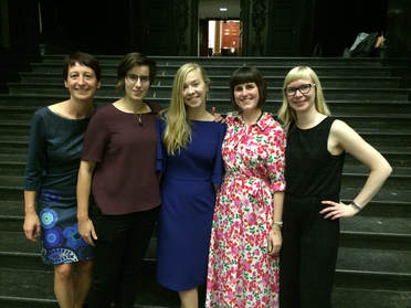
Miriam Bauwens defended successfully her PhD thesis on "Missing heritability in ABCA4-associated disease" on the 19th of September 2018 in Ghent. Congratulations Miriam!
Check out our latest paper published in Genetics in Medicine. You can read the abstract below, for the full text click here.
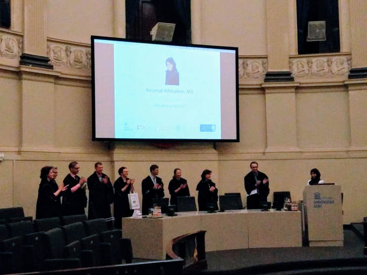
Purpose: Disorders or differences of sex development (DSDs) are rare congenital conditions characterized by atypical sex development. Despite advances in genomic technologies, the molecular cause remains unknown in 50% of cases. Methods: Homozygosity mapping and whole-exome sequencing revealed an ESR2 variant in an individual with syndromic 46,XY DSD. Additional cases with 46,XY DSD underwent whole-exome sequencing and targeted next-generation sequencing of ESR2. Functional characterization of the identified variants included luciferase assays and protein structure analysis. Gonadal ESR2 expression was assessed in human embryonic data sets and immunostaining of estrogen receptor-β (ER-β) was performed in an 8-week-old human male embryo. Results: We identified a homozygous ESR2 variant, c.541_543del p.(Asn181del), located in the highly conserved DNA-binding domain of ER-β, in an individual with syndromic 46,XY DSD. Two additional heterozygous missense variants, c.251G>T p.(Gly84Val) and c.1277T>G p.(Leu426Arg), located in the N-terminus and the ligand-binding domain of ER-β, were found in unrelated, nonsyndromic 46,XY DSD cases. Significantly increased transcriptional activation and an impact on protein conformation were shown for the p.(Asn181del) and p.(Leu426Arg) variants. Testicular ESR2 expression was previously documented and ER-β immunostaining was positive in the developing intestine and eyes. Conclusion: Our study supports a role for ESR2 as a novel candidate gene for 46,XY DSD. On Friday December 8 2017 Basamat ALMOALLEM, a Saudi ophthalmologist in training, successfully obtained her PhD in Health Sciences on the basis of her doctoral thesis ‘Unraveling the molecular basis of genetically heterogeneous developmental eye disorders’. Her findings unveiled examples of care valued by both patients and clinicians in the field of ophthalmic genetics. She has taken great steps towards tackling some of the challenges ophthalmic genetics is facing today.
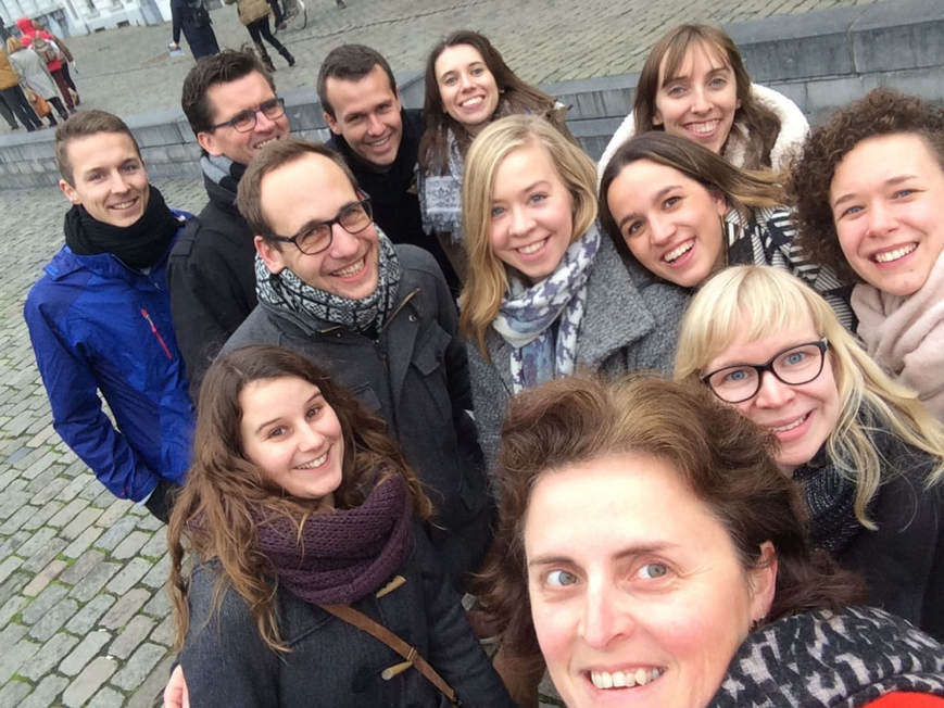
For our new team website we obviously needed a nice team picture. The crime scene of choice was the historic centre of Ghent. We would like to thank our photographer Pieter-Jan Volders for the beautiful photos. In the picture below you can see the models getting ready for the shoot.
|